Antique Microscopes: A Great Hobby!
Robert Brodell, MD, Chair of the Department of Pathology, was the first dermatopathology fellow for Daniel J. Santa Cruz, MD, an internationally acclaimed expert in dermatopathology. When Dr. Santa Cruz passed away on March 30, 2020, Dr. Bob inherited the antique microscope collection. Microscopes are becoming obsolete as inexpensive scanners, high-resolution computer screens, and computer joy sticks become commonplace. Still, these antique microscopes can be seen as sculptures commemorating key periods during the history of medicine as seen in Figure 1. Their key role will never be forgotten as we stand on the shoulders of the great physicians and scientists who used these instruments.
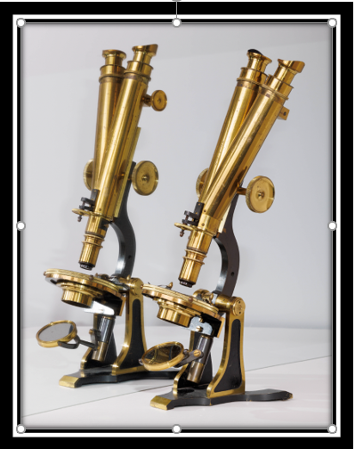
Figure 1: This early brass, binocular, single-objective microscope has a cast brass base with reverse Y-claw foot design and carrying two flat, cast pillars. The base and pillars are black Japanned except for the feet and a narrow bright strip along the edges. The lister limb is also blackened and is secured to the stand with trunions. The body tubes are attached to the limb by a rack and pinion running in a Jackson groove. This course focus system is operated by double hand wheels while fine focus utilizes a screwthread design with a thumbwheel acting on a sprung lever in the nosepiece. The main body tube is vertical with the binocular tube inclined to the left. It uses a Wenham prism. The intra-ocular distance can be increased by extending the nose pieces (H Crouch, 51 London Wall, London #573 circa 1870).
The Santa Cruz Antique Microscope Collection will be displayed in a series of exhibits in the glass cabinet adjacent to the front door of the pathology suite. Three microscopes are on display now. 3 more will go on display May 1, 2023.